THE MICHIGAN DAILY SUNDAY,
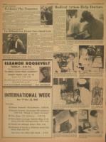
SD:Medical Artists Help Doc
m imen Pl q Tam arnaw
NOVEMBER 16, 19
tors
To comply with requests from
doctors at University Medical Cen-
ter who want illustrations of medi-
cal techniques, various parts of
the human anatomy and operating
procedure, the University has es-
tablished one of the largest medi-
cal illustration departments in the
United States.
Located in a corner of the sub-
basement of University Hospital,
four illustrators and two drafts-
men draw and copy everything
from eye corneas to aortic arches.
Doctors use these illustrations for
teaching aids in the Medical
School. -
Prof. Gerald P. Hodge, head of
the department, said illustrations
are often better than photographs,
for there is no extraneous material
which prevents an accurate pic-
ture of the specimen, particularly
in the operating room.
I"To become an illustrator, it is
necessary to take a three year
graduate course in medical illus-
tration," Prof. Hodge said.
Although medical illustration is
fairly new to most people, the pro-
fessor said one of the early ex-
amples was found in Egypt show-
ing Cleopatra giving birth to her
children.
The father of modern medical
illustration is Leonardo DaVinci
and it was first begun in the
United States by Max Brodel in
1890.
Prof. Hodge added that medical
illustration at the University
started in 1925,tbut an actual de-
partment was established only
three years ago.
OPERATING ROOM-Medical illustrators are often found in the. operating room where doctors
want illustrations of special operating techniques. Drawings showing the use of heart oxygenating
pumps which supply oxygen to the heart during cardiac surgery and pictures of an aortic transplant
are just some types of illustrating these artists do. Doctors then use these drawings for instruction
in the classrooms.
SLIT LAMP-An important job for the illustrators is to draw a
part of the human anatomy under actual conditions. The cornea of
the eye is being studied using this machine which sends a beam
of light, which may be focused on any part of the eye, onto the
cornea."
BOOK DISPLAY-Several projects of the medical illustrators are
the illustrations of textbooks. Mary Lou Cummings, one of the
artists, shows some of the types of illustrations used in the books.
Including pictures of body organs, nervous system and the muscles
of the body.
IN THE WARD-Prof. Gerald P. Hodge, head of the department of medical illustration, often works
in the wards of University Hospital. Doctors request illustrations of various disease formations for
study and for visual aids in teaching medical students how to recognize the most simple to the most
complex lesions and markings. Visual aids instruction is one of the most important parts of a
medical student's education.
MODEL - Skeletons are often
used by the illustrators when
they draw pictures of the skele-
tal system.
_ (
' .
, ?
Scanned image of the page. Keyboard directions: use + to zoom in, - to zoom out, arrow keys to pan inside the viewer.
November 16, 1958 (vol. 69, iss. 53) - Image 10
- Resource type:
- Text
- Publication:
- The Michigan Daily, 1958-11-16
Disclaimer: Computer generated plain text may have errors. Read more about this.