health & wellness
Local physicians find 3D-printed models helpful in surgery, treatment.
I
Ruthan Brodsky
Contributing Writer
A
dvances in medical technology
have significantly changed the
world of medical imaging in
the last decade. Two-dimensional imaging,
such as magnetic resonance imaging (MIU),
computer tomography (CT scan) and ultra-
sound, have been around since the early
1970s and provide a fairly good image of a
patient's internal organs.
Although the image resolution of these
devices has improved greatly over the years,
the technology itself remains fixed.
In the early 1980s, 3D printing was
introduced and was quickly used by the
manufacturing industry to produce product
prototypes, creating models and molds.
Instead of using ink to print text and pic-
tures, 3D printers lay down layer after layer
of plastic or some other flexible material
to build a physical model of the object.
Just last year, this technology made head-
lines when the crew of the International
Space Station used transmitted digital
plans to print a wrench from a 3D printer
onboard the space shuttle. Computer-aided
design (CAD) was used to draw up plans
that were relayed to the space station, where
it took four hours to print the finished
product.
Today, medical applications for 3D print-
ing are expanding rapidly and are expected
to revolutionize health care in the next
decade.
"In previous years, the major focus of
cardiology and cardiac surgery was coro-
nary artery disease and its treatment:' says
Dr. Adam Greenbaum, M.D., Farmington
Hills, co-director of the Henry Ford Center
for Structural Heart Disease and past
director of the cardiac catheterization lab
at Henry Ford Hospital. "Today the field
of treating structural heart disease has
exploded:'
Structural heart disease often refers to
congenital (birth) heart defects or condi-
tions that develop later in life and may
include abnormalities of the valves and
chambers of the heart wall, both of which
interrupt the natural flow of blood through
the heart.
"Over the last five years, we've been
using minimally invasive heart surgery to
close holes and to repair and replace valves
using catheters to deliver these treatments
through very small incisions:' Greenbaum
says. "Using a combination of imaging soft-
ware, we can create a replica of the patient's
heart and of the area to be treated, which
helps the surgeon become familiar with the
patient's anatomy. Using the digital data
from the imaging [MRI and CT scan], a
3D-printed replica or model of the valves
and heart is created and used to plan the
transcatheter surgery.
"This model enables us to become
familiar with the patient's anatomy, and
we have a better chance of anticipating any
unforeseen issue before we even start the
procedure Greenbaum says. "The model
also helps us decide which heart valve size
to use for the patient. With CAD and 3D
printing, we can easily predict the size and
fit of the valve.
As a result, these models have increased
the safety of structural heart surgeries
because we're able to plan a more correct fit
and proper placement of the valve. Using
the replica also makes it easier to help the
patient and family better understand the
procedure'
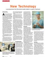
Dr. Adam Greenbaum, co-director of the Henry
Ford Center for Structural Heart Disease, says 3D
replicas of a patient's heart can help doctors plan
surgeries more efficiently.
48
May 28 • 2015
3D Printing History
3D printing is an additive manu-
facturing process that uses a digi-
tal model to create a 3D object by
using layering techniques with
some type of material. A sophis-
ticated computer-aided design
(CAD) program determines how
these layers of a material are laid
down.
The technology dates back
about 30 years and has been
used extensively in the auto and
aerospace industries to help pro-
duce prototypes for new cars and
car parts and lighter versions of
complex parts for airplanes.
In the healthcare area, 3D
printing has had a major impact
on hearing aid manufacturing
because everyone's ear canal is
shaped differently. The use of 3D
printing allows custom-shaped
devices to be produced efficiently
and cost effectively. For instance,
the outer shell of in-the-ear hearing
aids is likely to be 3D printed. As a
result, about 10 million 3D-printed
hearing aids are in circulation.
If you have a dental implant,
there's even a bigger chance that the
implant was also 3D printed. U.S.
dental labs have invested in technol-
ogy that can scan a patient's teeth so
that new teeth can be produced just
by pressing a button. Today, dentists
are increasingly creating implants
made of durable plastic or medical
ceramics using this technology.
Orthopedics
"Creating relatively inexpensive
images and models of a patient's
unique anatomy helps surgeons bet-
Orthopedic surgeon Dr. Robert Kohen holds
ter visualize complex fractures and
some custom 3D surgical models.
deformities," says Robert Kohen,
M.D, Bloomfield Hills, orthope-
dic surgeon Beaumont Hospital.
preoperatively determine the exact size and
"Physicians generate digital files from clini-
alignment of the patient's knee before sur-
cal data to make custom surgical models.
gery. This data can be used to create custom
They can use the model for presurgical
guides or implants to improve the surgery:'
guidance or use it in the operating room as
According to speakers at the 2014 Inside
3D Printing Conference and Expo in New
a guide for the surgery:'
The ability to produce 3D images and
York, advances in 3D printing and medical
even custom implants helps to resolve the
technology will soon make it possible to
problem in orthopedics when standard
construct human tissue in a lab, implant it
implants don't work for some patients:'
in a patient and watch it grow in the body.
Kohen says. "For example, the 3D models
Tissue engineering, as it's called, is one of
help surgeons determine the safest way to
the new technological advances researchers
perform the surgery and where to precisely
and doctors have made in the medical 3D
printing field.
position the replacement. They are also
used during pre-operative planning for
challenging surgical cases.
The Future
"By utilizing 3D-formatted images, sur-
Medical visualization is the use of comput-
geons can better comprehend the character
ers to create 3D images from medical imag-
of a complex fracture even before they
ing data sets. It's a young science discipline
begin surgery," Kohen says. "This ability,
relying on advances in computing.
RealView Imaging LTD in Israel recently
of course, increases the precision of the
surgery, which means patients have a better completed a successful clinical study in
chance of an excellent outcome:'
which surgeons used live-action 3D holo-
Another major benefit for using 3D mod- grams of their patient's beating heart to help
eling is that very personalized joints can be
them operate. The system gives surgeons
ordered and constructed.
information about the entire organ in real
Surgeons have choices of many possible
time, an important factor in surgery.
knee sizes during surgery and select the
Recently, imaging techniques have been
one that appears to be the closest fit:' says
developed that work on the level of mol-
Kohen. "They precisely shape the bone until ecules and genes. The potential comes in
the implant fits perfectly. Improper sizing
identifying pathological processes at an
can lead to stiffness and inferior outcomes.
early stage before they become apparent in
Using a CT scan or MRI, the surgeon can
the form of tumors.
❑